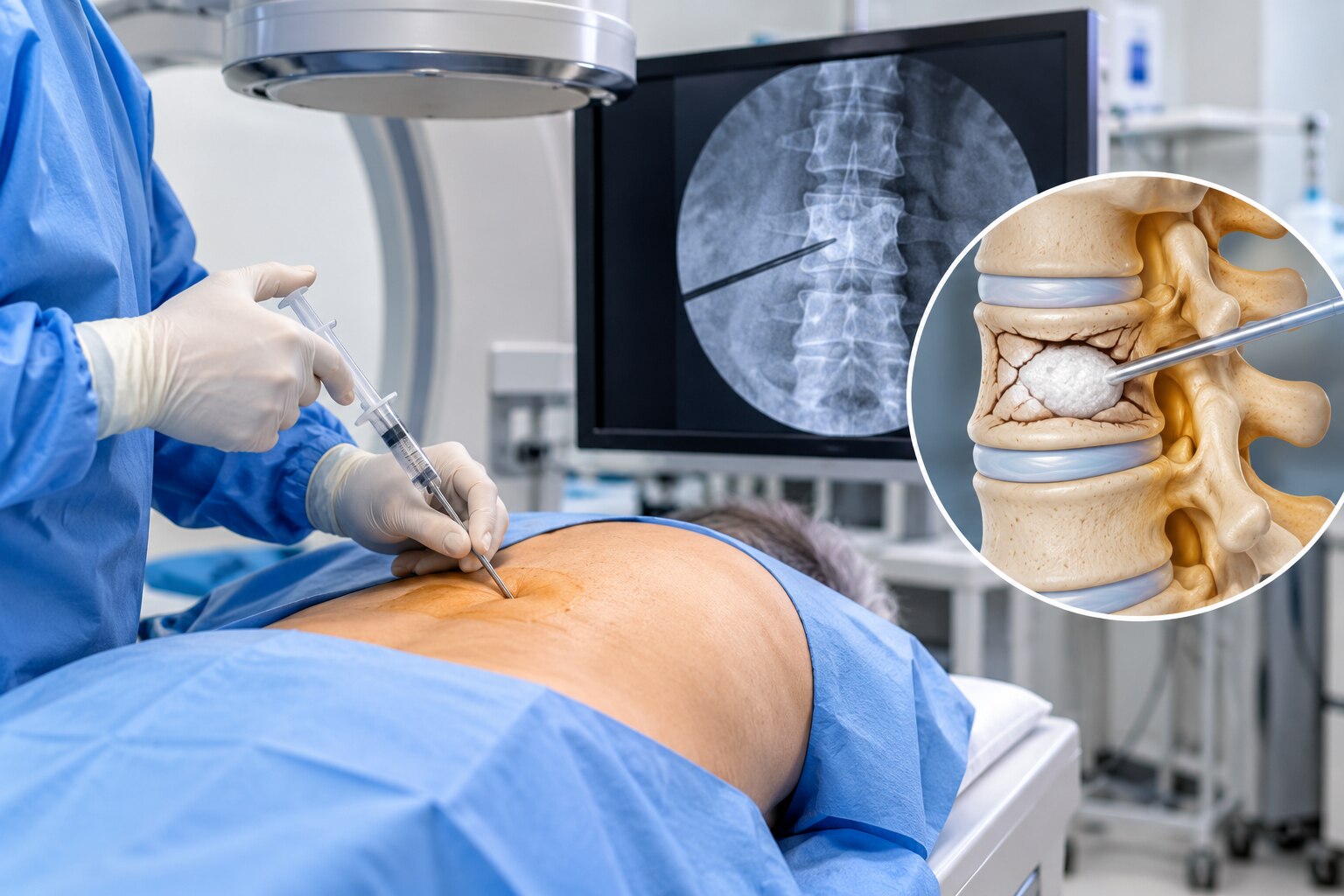
A vertebral compression fracture can cause sharp, focal pain that worsens when you stand, walk, cough, or change position, but an abnormal X-ray does not prove that the fracture is causing today’s symptoms. By the end, you will understand how clinicians confirm an active fracture, how vertebroplasty stabilises it, and when its potential benefit justifies the procedural risks.
Key takeaways
- Confirm the painful level with examination and MRI or other imaging.
- Vertebroplasty stabilises a fractured vertebra with bone cement.
- Consider vertebroplasty after persistent pain despite appropriate conservative care.
- Compare cement leakage, height restoration, and other risks with kyphoplasty.
How can you tell whether a compression fracture is causing the pain?
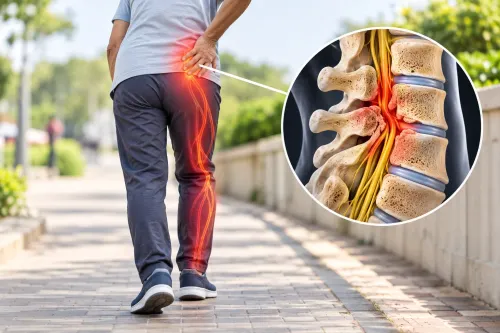
Vertebral compression fracture pain is usually sudden, focal back pain that begins after a fall, lifting effort, cough, or ordinary movement in a fragile spine. It centres over one vertebral level, worsens when you stand, walk, twist, or change position, and often improves when you lie still.
Pressing directly over the affected bone may reproduce the pain.
Clinicians look for patterns that point elsewhere:
- Muscular back pain is usually broader, linked to muscle spasm or a strained movement, and tender in the muscles beside the spine rather than directly over one vertebra.
- Disc pain often produces central low-back pain with buttock or leg symptoms; numbness, tingling, or weakness suggests nerve-root irritation rather than pain confined to one spinal level.
- Spinal stenosis usually causes leg pain, heaviness, or weakness while standing or walking, with relief from sitting or bending forward.
- Chronic osteoporotic aching is diffuse and persistent, without a clear recent change or sharply tender level.
- An old healed deformity may appear on an X-ray but cause no current pain.
MRI helps decide whether the fracture is active. Bone-marrow edema on fluid-sensitive or fat-suppressed images supports a recent or still-painful fracture; its absence supports an old, healed deformity. MRI also checks for infection, cancer, canal compromise, disc disease, or a non-spinal pain source.
A fracture on an old X-ray alone does not prove it is causing today’s symptoms.
What happens during vertebroplasty, and how can cement reduce pain?
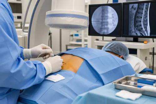
You lie face down on a procedure table so the affected vertebra is accessible from your back. After skin cleaning and sterile draping, the team uses local anaesthetic, often with medication for relaxation; general anaesthesia is used when necessary.
The vertebroplasty procedure follows a controlled sequence:
- Imaging identifies the target level and safest needle path.
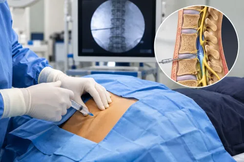
- Fluoroscopy and CT guidance direct a narrow needle through the pedicle or directly through the vertebral body.
- The clinician confirms the needle tip lies inside the fractured vertebra.
- Liquid polymethylmethacrylate PMMA cement is injected gradually and watched on live images.
The cement thickens inside the porous bone over minutes. Once hardened, it creates an internal support structure that reduces small movements between fragments and within cracks in the vertebral body. Those movements can trigger pain when you stand, turn, cough, or walk, so vertebral fracture stabilisation may reduce mechanically driven pain.
Vertebroplasty does not rebuild crushed bone, restore lost vertebral height, correct kyphosis, or treat osteoporosis. Its purpose is stabilisation, not structural reconstruction. The operator stops or changes the injection if cement approaches the spinal canal, nerve openings, or veins, because leakage into those spaces can cause neurological or circulatory complications.
Careful CT or MRI review helps define that risk before the needle enters.
Which patients are considered for vertebroplasty instead of continued conservative care?
Vertebroplasty is considered when severe, focal pain from a recent compression fracture remains disabling despite appropriate conservative treatment. The pain should match the fractured level, with local tenderness and imaging evidence that the fracture is active rather than an old deformity.
- MRI bone-marrow oedema on fluid-sensitive or fat-suppressed sequences supports a recent, painful fracture.
- CT or X-ray should show a compression fracture that matches the MRI and examination.
- The clinician should exclude infection, cancer, spinal canal compromise, and pain from a disc, muscle, hip, or another source.
- Pain should remain substantial despite medication, safe activity modification, osteoporosis care, and a suitable period of recovery.
| Option | What it offers | When it applies |
|---|---|---|
| Analgesics and activity modification | Controls symptoms while the fracture settles; prolonged bed rest causes weakness and clot risk | Initial care for most uncomplicated fractures |
| Short-term brace | Limits painful movement but can restrict breathing or weaken trunk muscles if overused | Selected patients during early recovery |
| Osteoporosis treatment and time | Reduces future fracture risk; many fractures improve over several weeks | Essential whether or not augmentation is performed |
| Vertebroplasty | Stabilises the painful vertebral body without rebuilding bone or treating osteoporosis | Persistent, severe, imaging-confirmed pain after conservative care |
The VERTOS II trial reported greater relief in a narrowly selected group with acute fractures, MRI oedema, pain of at least 5/10, and symptoms lasting up to six weeks. By contrast, 2009 sham-controlled trials found no significant short-term advantage in broadly selected patients. Those findings support selective use, not routine treatment for every painful fracture.
How does vertebroplasty compare with kyphoplasty, and what risks must you weigh?
Vertebroplasty versus balloon kyphoplasty comes down to the goal: PMMA cement injection alone stabilizes the fracture, while kyphoplasty inflates balloons first and then fills the created cavity with cement. Balloon inflation can improve vertebral height restoration and reduce kyphotic angulation, but it cannot reliably rebuild a severely collapsed vertebra.
| Option | Main step | Practical trade-off |
|---|---|---|
| Vertebroplasty | Needle placement followed by PMMA injection | Uses less equipment and often has lower procedural cost; it does not restore height |
| Balloon kyphoplasty | Balloon inflation followed by cement injection | Requires balloons and an inflation system, adding equipment, access steps, and cost; it may restore height |
Both procedures carry cement leakage risk. Leakage into the disc or nearby soft tissue is common on imaging and usually causes no symptoms, but leakage into the spinal canal can compress a nerve, while venous leakage can cause pulmonary cement embolism. Infection and bleeding risk also require screening, alongside rare cardiopulmonary complications.
Choose another diagnosis or treatment first when:
- MRI or CT suggests tumour, epidural disease, or pain unrelated to the fracture; malignancy needs oncologic assessment, and cement does not treat the tumour.
- Retropulsed bone or epidural material causes weakness, numbness, or bladder and bowel changes; surgical decompression may be necessary.
- Active infection, uncorrected coagulopathy, material allergy, or inability to lie prone makes augmentation unsafe.
- Severe osteoporosis remains untreated; medication, fall prevention, and fracture-risk management are still necessary after either procedure.
What should assessment and follow-up include before and after augmentation?
MRI or CT review must confirm that the painful level is active and safe to treat. MRI fluid-sensitive or fat-suppressed images showing bone-marrow oedema support an acute fracture; absent oedema points towards a chronic fracture or healed deformity. CT shows posterior-wall defects, canal narrowing, or a fracture pattern that increases cement-leakage risk.
- Record focal tenderness, pain severity, mobility, and a neurological examination of strength, sensation, reflexes, and bowel or bladder function.
- Review anticoagulants, antiplatelet drugs, platelet count, clotting results, diabetes control, fever, skin infection, and other bleeding or infection risks.
- Use imaging and history to investigate spinal tumour and infection, especially with cancer history, unexplained weight loss, fever, night pain, or abnormal bone destruction.
- Check whether hip, rib, sacroiliac, muscular, disc, or nerve pain is actually causing the symptoms. A specialist pain assessment prevents cement treatment being aimed at the wrong structure.
After augmentation, arrange follow-up for wound problems, fever, worsening pain, new weakness, numbness, breathing difficulty, or bladder and bowel changes. Confirm functional improvement rather than judging success only by a scan.
Osteoporosis treatment after vertebroplasty remains essential: discuss bone-density testing, calcium and vitamin D intake, prescribed anti-osteoporosis medication, strength and balance exercise, vision review, and fall hazards at home. Ask the specialist:
- Is the fracture acute, chronic, or uncertain?
- What finding proves this level causes my pain?
- What are my cement-leakage and bleeding risks?
- What is the plan if pain persists?
- Which osteoporosis treatment and fall-prevention steps do I need?
Spine And Pain Management Centre can use these questions to structure a consultation around diagnosis, procedural risk, and long-term fracture prevention.
Related service
Pain Treatment ₹700 Dr Prakash Deshmukh at Spine and Pain Management Centre in Kalyan provides advanced Vertebroplasty Treatment for patients suffering from spinal... View service → |
Frequently asked questions
How can you tell whether a compression fracture is causing the pain?
Doctors match sudden, focal pain and tenderness with examination and imaging. MRI can show bone-marrow oedema, which helps identify a recent painful fracture.
What happens during vertebroplasty, and how can cement reduce pain?
Under imaging guidance, a doctor places a needle through the skin into the fractured vertebra and injects bone cement. The hardened cement stabilises the vertebra, which can reduce movement-related pain.
Which patients are considered for vertebroplasty instead of continued conservative care?
Doctors consider it when imaging confirms a painful, recent compression fracture and significant pain persists despite appropriate medicines, activity changes, and other conservative treatment.
How does vertebroplasty compare with kyphoplasty, and what risks must you weigh?
Both procedures inject cement to stabilise a fractured vertebra. Kyphoplasty first uses a balloon, which can restore some vertebral height; both carry risks including cement leakage, infection, bleeding, nerve injury, and new fractures.
What should assessment and follow-up include before and after augmentation?
Assessment should review fracture age, imaging, bone strength, neurological symptoms, medicines, and other pain causes. Follow-up checks pain, mobility, wound problems, neurological changes, cement-related complications, and osteoporosis treatment.