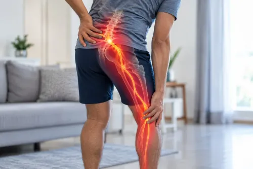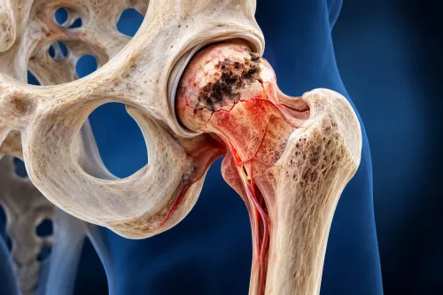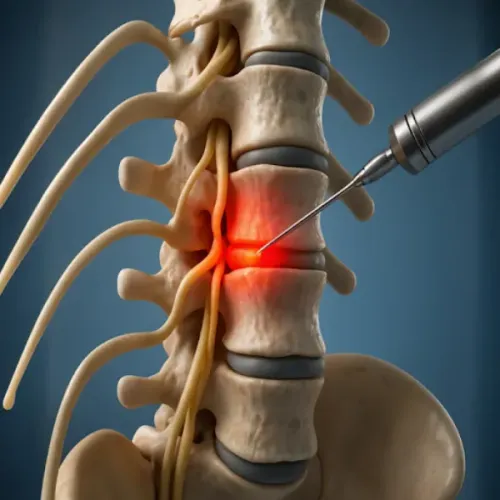
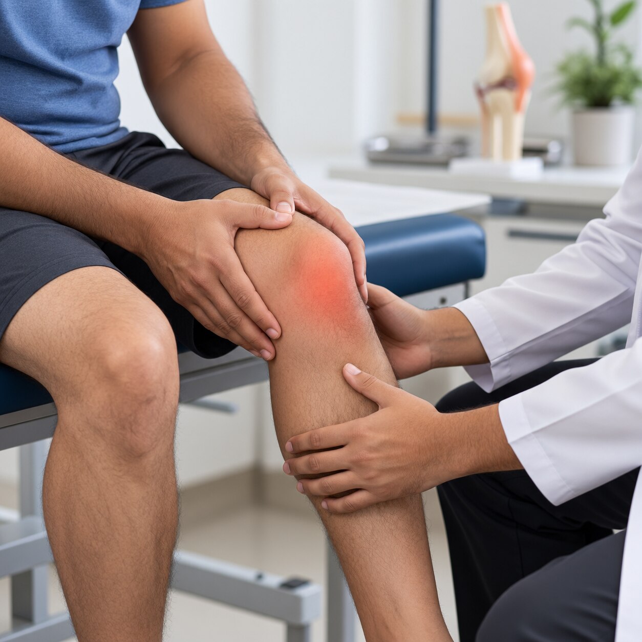
Knee pain can come from the joint itself, a ligament, tendon, meniscus, kneecap, hip, nerve, or lower spine, so the most painful spot does not always identify the cause. By understanding the history, examination, tests, and warning signs clinicians use, you can ask better questions and see why one treatment fits your findings while another does not.
Key takeaways
- Record pain location, onset, duration, severity, and whether it travels.
- A hip, nerve, or spine examination can reveal non-knee causes.
- Use X-rays, ultrasound, or MRI when examination findings require more evidence.
- Blood tests or joint aspiration help investigate infection, gout, or inflammation.
What Your Symptom History Reveals Before the Examination
A careful knee pain history helps identify whether the problem began suddenly after trauma or developed gradually through degeneration, overuse, inflammation, or referred pain. Record the pain location with one finger, its duration, severity, and whether it travels from the hip, thigh, or lower back. Pain intensity alone does not measure structural damage.
- Describe the injury mechanism: a twist, direct blow, fall, pivot, sudden stop, or increase in running can point toward different tissues.
- Note what triggers symptoms, such as stairs, squatting, walking, rest, or turning in bed, and whether the knee gives way, catches, or remains locked.
- Describe knee swelling: immediate swelling after injury suggests bleeding into the joint, while swelling that appears hours later can follow irritation; a hot, rapidly swollen knee with fever needs urgent assessment.
- Report morning stiffness, including whether it lasts a few minutes or more than 30 minutes, and mention psoriasis, gout, autoimmune disease, infection, or previous surgery.
| Symptom pattern | What it may suggest | What changes urgency |
|---|---|---|
| Gradual pain with activity | Osteoarthritis, tendon, or kneecap disorder | Function declining or pain persisting |
| Twist followed by swelling or instability | Meniscus or ligament injury | Persistent giving way or inability to walk |
| Hot swollen knee with fever | Infection or crystal arthritis | Same-day medical assessment |
| Persistent inability to fully extend | True locked knee | Prompt orthopaedic assessment |
How the Physical Examination Separates Knee, Hip, Nerve, and Spine Causes
A knee physical examination starts with your gait, posture, and ability to bear weight, not just the painful spot. The clinician compares both legs, checks alignment, muscle wasting, swelling or effusion, warmth, and bruising, then measures range of motion while you bend and straighten the knee.
- Joint-line tenderness points toward a meniscus or arthritis; pain directly over the patellar tendon, quadriceps tendon, or bursae suggests a soft-tissue source.
- Tracking of the kneecap, compression pain, and tenderness around the patella help identify patellofemoral problems.
- Knee stability tests, including Lachman, anterior and posterior drawer, and varus-valgus stress tests, assess the anterior cruciate, posterior cruciate, and collateral ligaments.
- Squatting, twisting, and McMurray or Thessaly manoeuvres can reproduce meniscal pain, catching, or clicking.
- Hip range of motion, especially internal rotation, is checked because hip arthritis or other hip disease can refer pain to the knee.
- Strength, reflexes, sensation, and straight-leg raising help detect femoral or sciatic nerve involvement.
- Lumbar movement and a neurologic examination look for a pinched nerve or lumbar spine disorder producing pain, tingling, or weakness below the hip.
A hot, swollen knee with fever or marked illness needs urgent assessment for infection. Examination alone cannot exclude septic arthritis; aspiration for cell count, Gram stain, culture, and crystal analysis is often necessary. Pain without local knee findings makes a referred hip, nerve, or spine source more likely.
When X-Rays, Ultrasound, or MRI Add Useful Evidence
A weight-bearing knee X-ray is usually the starting image for persistent knee pain when arthritis, alignment change, or previous injury is suspected. Standing views show joint-space narrowing and load-related alignment more clearly than a non-weight-bearing image.
The result cannot predict pain by itself: severe X-ray changes may cause little discomfort, while modest changes can be very painful.
| Test | Useful evidence | When it applies |
|---|---|---|
| Weight-bearing knee X-ray | Bone shape, joint-space narrowing, alignment, fractures, and osteoarthritis changes | Persistent pain, suspected arthritis, deformity, or injury |
| Knee ultrasound | Fluid, superficial tendons, bursae, and guidance for aspiration or injection | Swelling, suspected tendon or bursal problems, or a procedure needing precise placement |
| Knee MRI | Menisci, ligaments, cartilage, bone marrow, and deeper structural injury | Persistent symptoms after examination and X-ray, suspected internal derangement, or an occult injury |
A knee ultrasound is operator-dependent and cannot replace an X-ray for alignment or a knee MRI for many deep injuries. MRI findings also need restraint: degenerative meniscal tears and cartilage defects are common without symptoms.
A reported meniscus tear matters when its location matches the pain, examination findings, swelling, or true mechanical locking; intermittent clicking alone does not prove that surgery is needed. Hip movement, gait, and spine and nerve findings must also be considered because pain felt at the knee can start elsewhere.
When Blood Tests or Joint Aspiration Are Necessary
A hot, swollen knee with fever, chills, or feeling acutely unwell needs urgent assessment for septic arthritis, because infection can rapidly damage the joint. Blood tests such as a complete blood count, C-reactive protein, erythrocyte sedimentation rate, and blood cultures support the assessment, but normal results cannot reliably exclude infection.
A knee joint aspiration removes synovial fluid through a needle for direct testing. Ask what the sample will assess:
- Cell count and differential: measures the intensity and type of inflammation.
- Gram stain and culture: looks for bacteria and guides antibiotic choice.
- Crystal examination under polarized light: identifies monosodium urate crystals in a gout knee or calcium-pyrophosphate crystals in pseudogout.
Synovial fluid analysis matters because a serum urate result alone does not confirm gout; urate can be normal during an acute flare. Crystal identification also separates gout from calcium-pyrophosphate deposition, which requires a different clinical assessment.
Persistent warmth, swelling, prolonged morning stiffness, or several painful joints raises concern for inflammatory arthritis. Clinicians may order rheumatoid factor, anti-CCP antibodies, and inflammatory markers, but these tests are not general screening tools and a negative result should not delay referral when the examination suggests rheumatoid arthritis.
Do not proceed with an injection until infection has been considered and, when needed, excluded.
How Evaluation Findings Shape the Treatment Decision
The next step depends on the pain generator, not the scan alone. Degenerative, traumatic, inflammatory, infectious, referred, and nerve-related pain require different risk-benefit decisions. Pain intensity does not reliably match arthritis damage, so function, examination findings, duration, and patient preferences matter more than an X-ray grade by itself.
- Short-lived pain after a clear injury, with knee instability, major swelling, or inability to bear weight, may require urgent assessment, bracing, rehabilitation, or an orthopaedic referral.
- Activity-related pain lasting months, with stiffness of 30 minutes or less in an adult aged 45 or older, often supports conservative care such as exercise, strength training, weight management, and symptom-directed medication before invasive treatment.
- Mechanical knee symptoms such as true locking, catching, or a displaced injury prompt closer assessment for meniscal, loose-body, or cartilage problems; an MRI finding alone does not justify arthroscopy.
- Age, work demands, sport, walking distance, and the activity you want to regain determine whether the plan prioritises rehabilitation, pain relief, joint-preserving procedures, or replacement discussion.
- Diabetes, blood-thinning medication, infection risk, heart disease, kidney disease, and previous operations alter medication and procedure safety.
- Failure of an adequate prior treatment trial can justify changing strategy, not repeating it unchanged. A diagnostic injection may identify a pain source, but temporary relief does not guarantee lasting procedural benefit.
Spine And Pain Management Centre can use this decision framework to separate a knee source from referred pain before offering an intervention. That distinction prevents a treatment aimed at the wrong structure.
Related service
Pain Treatment ₹700 Knee pain can affect mobility, daily activities, and overall quality of life. At Spine and Pain Management Centre in Kalyan, Dr. View service → |
Frequently asked questions
What should you record before a knee pain evaluation?
Record where the pain occurs, when it began, how long it has lasted, its severity, and whether it travels from the hip, thigh, or lower back. Note injuries, overuse, swelling, stiffness, locking, instability, fever, and treatments already tried.
How can a physical examination identify a non-knee cause?
The clinician compares both knees, checks movement, swelling, tenderness, stability, strength, and gait, then examines the hip, nerves, and lower back. Pain reproduced by hip or spine movements can indicate referred pain rather than a knee disorder.
When are X-rays, ultrasound, and MRI used for knee pain?
X-rays show bone alignment, fractures, and joint-space narrowing. Ultrasound can assess fluid, tendons, and accessible soft tissues while guiding procedures. MRI provides detailed images of cartilage, menisci, ligaments, bone marrow, and other internal structures when those findings would change treatment.
When are blood tests or joint aspiration necessary?
Blood tests help investigate infection, inflammatory arthritis, or gout when symptoms and examination suggest them. Joint aspiration removes synovial fluid for cell counts, crystal analysis, and culture, especially when a hot, swollen knee raises concern for infection or crystal arthritis.
How do evaluation findings determine the treatment approach?
Findings determine whether you need activity modification, exercise-based rehabilitation, medication, an injection or other minimally invasive treatment, further investigation, or urgent specialist care. Treatment should match the identified cause rather than pain intensity alone.