Avascular necrosis can begin with a blood-supply problem that is invisible on an X-ray, then progress to collapse of the femoral head and painful arthritis. By the end, you will know how staging, treatment planning, rehabilitation and measurable mobility goals determine whether a non-surgical regenerative program is a reasonable joint-preservation option.
Key takeaways
- Use MRI staging to detect AVN before collapse changes the treatment goal.
- Follow prescribed weight-bearing limits to reduce stress on weakened bone.
- Ask how the regenerative program will measure pain, function, and structural change.
- Joint preservation is more realistic before collapse or advanced arthritis develops.
What happens to the joint in avascular necrosis?
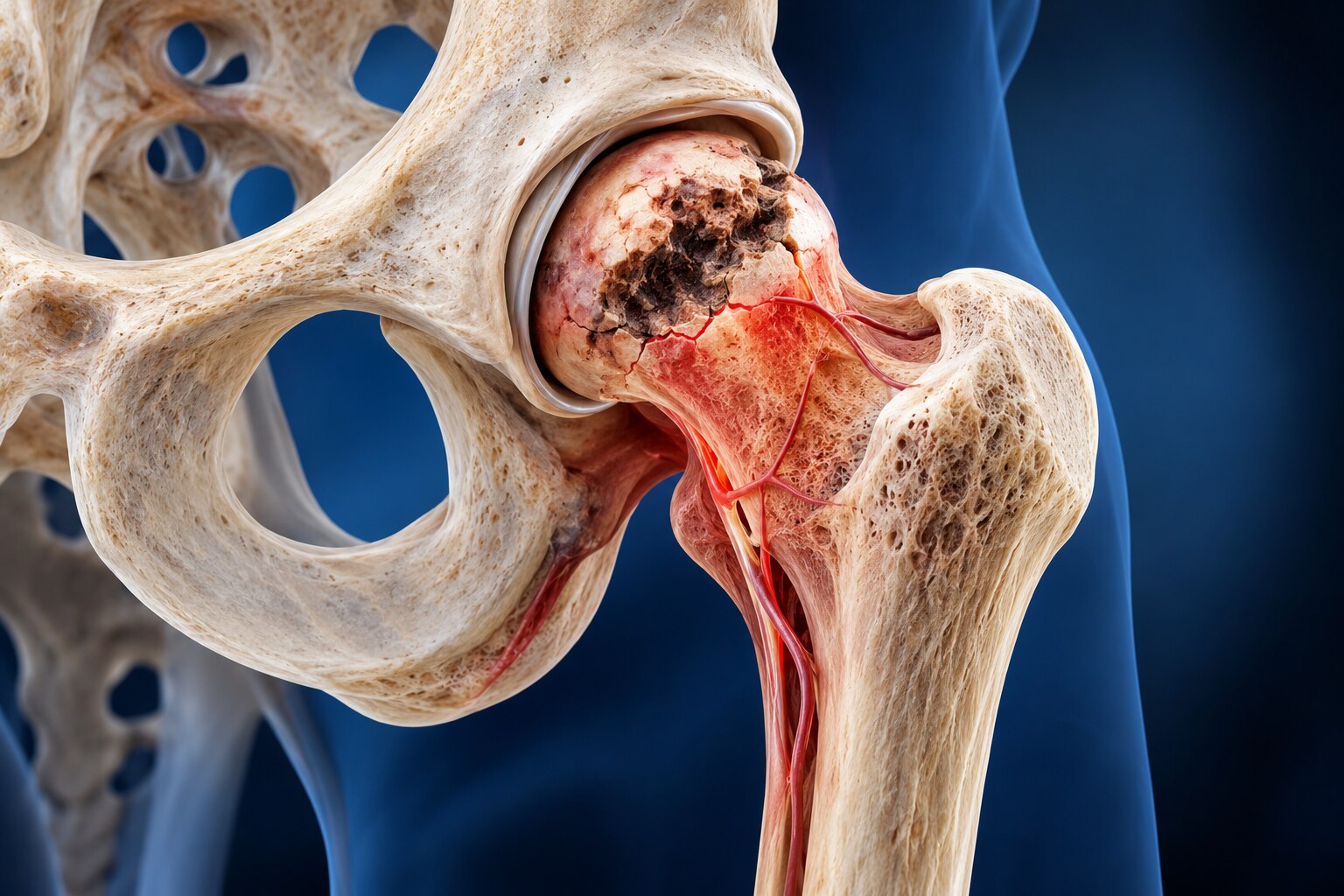
Avascular necrosis starts when loss of blood supply to bone deprives bone cells of oxygen and nutrients. The affected area dies, becoming weaker under normal loading. This process is also called osteonecrosis or AVN.
The hip is the most recognised site, particularly the rounded femoral head, but AVN also affects the knee, shoulder, ankle and small bones of the hands and feet. Hip disease can cause groin, buttock or thigh pain, restricted inward rotation, limping and reduced walking tolerance.
| Stage | What happens inside the joint | Common symptom pattern | Why it matters |
|---|---|---|---|
| Before structural collapse | Dead bone remains beneath an intact joint surface, although it is vulnerable to cracking and deformation. | No symptoms, activity-related pain, or aching with increasing walking. | Joint-preservation treatment has its strongest rationale before the surface caves in. |
| After structural collapse | The femoral head or another affected surface loses its round shape, altering load distribution and damaging cartilage. | Persistent pain, stiffness, a pronounced limp, difficulty standing or walking, and pain at rest. | Secondary arthritis can develop, making preservation harder and joint replacement more predictable. |
Symptoms alone cannot show whether collapse has begun. A person can walk better while the underlying lesion continues to weaken, and pain relief does not prove that dead bone has recovered. Imaging and follow-up therefore matter when assessing whether preservation remains realistic.
How do imaging and staging change the treatment decision?
The treatment decision turns on whether the femoral head is still structurally intact and whether the damaged area has a realistic chance of supporting the joint. Preservation is most plausible when AVN is detected before femoral-head collapse and advanced arthritis.
AVN diagnosis combines your pain pattern, walking tolerance, hip examination and imaging. Groin, buttock or thigh pain, restricted rotation and a limp raise concern, but symptoms alone cannot show how much bone is at risk. A joint can hurt less while the underlying lesion continues to progress.
- X-ray: It may look normal in early disease, then show density changes, a crescent-shaped fracture or femoral-head collapse.
- MRI for AVN: It detects earlier bone injury and maps the lesion’s size, depth and position before an X-ray becomes abnormal.
- Lesion location: Damage in a weight-bearing part of the femoral head carries greater mechanical risk than a small, non-weight-bearing lesion.
- Symptoms and examination: Pain, restricted movement and declining walking tolerance add urgency but do not determine stage by themselves.
- Cause and overall pattern: The clinician considers the underlying cause, the opposite hip and other affected sites when deciding how closely to monitor you.
A small, contained lesion without collapse may support load protection, surveillance and discussion of joint-preserving options. A large or weight-bearing lesion, visible surface failure, or established arthritis shifts the decision toward more predictable reconstructive treatment. Pain improvement after an injection does not prove that dead bone has recovered; follow-up imaging remains important.
What happens during a non-surgical regenerative program?
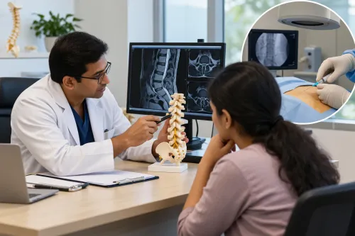
A regenerative program begins with a decision about suitability, not with an injection. The clinician reviews your symptoms, examination findings, X-rays and MRI, then weighs lesion size and location, collapse status, age, activity demands and the cause of AVN.
1. Consultation identifies the treatment goal, such as reducing pain, maintaining walking ability or preserving an early-stage joint. Record your medicines, steroid exposure, smoking history and other conditions because they affect risk and selection.
2. Imaging review confirms whether a non-surgical approach is reasonable. PRP and bone marrow concentrate are not interchangeable: preparation, cell content, dose and processing differ, and neither is proven to rebuild necrotic bone or prevent hip collapse.
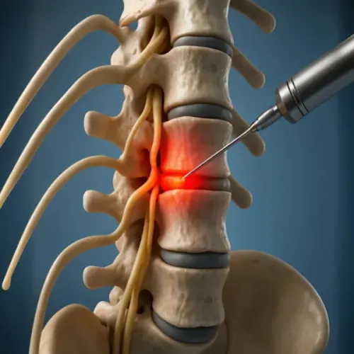
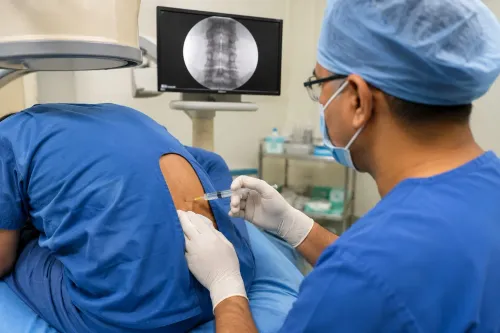
3. Injection planning matches the selected product to the target and uses imaging guidance when needed. The clinician explains preparation, anaesthesia, expected soreness, activity restrictions and risks, including bleeding, infection, nerve or tissue injury, contamination, immune reaction and failure to improve.
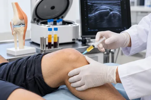
4. The procedure collects or prepares the material, then places it at the planned site. A pain flare can follow. Steroid injections require separate caution because repeated or systemic corticosteroid exposure is linked with AVN risk.
5. Follow-up checks pain, hip movement, walking tolerance and function, followed by rehabilitation instructions. Improvement does not prove that dead bone has recovered, so repeat imaging may be needed.
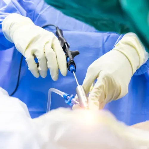
At Spine And Pain Management Centre, asking how the proposed product, target, imaging findings and outcome measures connect helps you assess a plan rather than accept a generic protocol. Core decompression is a separate joint-preserving operation for selected pre-collapse disease; after collapse or advanced arthritis, replacement is often more predictable.
Related service
Pain Treatment AVN Non-Surgical Regenerative Program Treatment ₹700 Dr Prakash Deshmukh at Spine and Pain Management Centre in Kalyan offers advanced AVN (Avascular Necrosis) Non-Surgical Regenerative Program... View service → |
How can you protect mobility while the joint is being assessed or treated?
Track function, not pain alone. Pain relief can improve walking without proving that dead bone has recovered or that collapse risk has ended. Use the same measures at follow-up:
- Walking tolerance: distance or minutes before pain forces you to stop
- Limp and stair ability
- Joint range, especially hip rotation
- Pain during activity and the following day
- Reliance on a cane, crutches or other support
Protected weight bearing reduces mechanical stress while the joint is vulnerable. Use a cane or crutches only with guidance matched to the affected joint, your balance and your strength; too much loading can aggravate symptoms, while unnecessary restriction can cause deconditioning.
Physiotherapy should preserve range without provoking joint pain, then build strength in the muscles supporting the joint. Low-impact exercise can maintain conditioning when running, jumping or long walks exceed your current tolerance. Increase distance and resistance gradually, and record any next-day flare.
Sleep supports rehabilitation by improving recovery and making pain easier to manage. Weight management reduces load across weight-bearing joints, while cause reduction means addressing the factor linked to your AVN, such as prescribed steroid exposure or alcohol use, with your clinician.
These steps support joint preservation but do not restore lost blood flow or reverse established collapse; pair them with scheduled clinical review and imaging when indicated.
How do you judge whether preservation is still a sensible goal?
Preservation remains a sensible goal when follow-up imaging shows no femoral-head collapse, the lesion is limited or favourably located, and symptoms remain manageable with protected loading and rehabilitation. Repeat X-ray or MRI, interpreted alongside examination and walking tolerance, matters because pain relief alone does not prove that dead bone has recovered.
Review the opposite hip when AVN risk factors or symptoms suggest bilateral disease.
| Option | Imaging and symptoms | What the decision means |
|---|---|---|
| Continue preservation | No collapse, stable lesion, tolerable symptoms and useful function | Continue load management, rehabilitation and carefully monitored treatment outcomes |
| Discuss core decompression | Early femoral-head AVN before substantial surface collapse, with persistent pain or a meaningful lesion | Seek a specialist opinion; this surgical option is not equivalent to a nonsurgical injection |
| Discuss another specialist option | Lesion size, location, age, activity demands or progression make one approach unsuitable | Compare joint-preserving procedures and their risks with observation |
| Consider joint replacement | Collapse, femoral-head deformity, joint-space loss or advanced arthritis with disabling pain | Hip replacement is often more predictable than attempting to regenerate a damaged surface |
Escalating pain, a new limp, reduced hip rotation or declining walking distance warrants reassessment rather than simply extending the same program. Once the surface has collapsed and arthritis limits the joint, joint replacement usually offers a clearer route to pain-limited function than preservation efforts.
Any clinician promising to preserve every joint should explain where that boundary lies.
Frequently asked questions
What happens to the joint in avascular necrosis?
Loss of blood supply kills bone cells, weakens the affected area, and can lead to structural collapse under normal loading.
How do imaging and staging change the treatment decision?
X-rays show shape and collapse, while MRI can reveal earlier disease. Stage, lesion size, location, symptoms, and joint-surface damage help determine whether preservation is realistic.
What happens during a non-surgical regenerative program?
A specialist reviews your diagnosis and imaging, sets activity and loading limits, and may combine targeted regenerative treatment with rehabilitation and follow-up measures.
How can you protect mobility while the joint is assessed or treated?
Use the prescribed walking aid or weight-bearing limit, choose low-impact movement, avoid painful loading, and complete exercises within the plan provided by your clinician.